Breaking news and analysis on politics, business, world national news, entertainment and more.
View Limbus Of Eye Location Pics
15/10/2020 00:00
View Limbus Of Eye Location Pics. This condition causes a milky white film to appear over your eyes and may change the way your eye coloring appears. The iris is the structure…
Biological Principals And Clinical Potentials Of Limbal Epithelial Stem Cells Springerlink from media.springernature.com
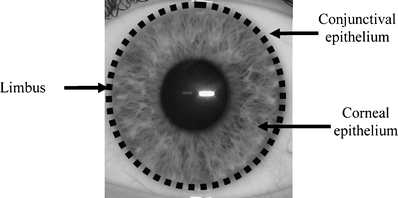
Ten layers of cells in the retina can be seen. Confocal images of the superior limbus (bottom left), nasal limbus (bottom middle), and inferior limbus (bottom right) show the presence of normal limbal epithelial cells. Limbal stem cells, also known as corneal epithelial stem cells, are stem cells located in the basal epithelial layer of the corneal limbus.they form the border between the cornea and the sclera.characteristics of limbal stem cells include a slow turnover rate, high proliferative potential, clonogenicity, expression of stem cell markers, as well as the ability to regenerate the entire corneal.
The retina is the part of the eye that receives the light and converts it into chemical energy.
Since rabbits open their eyes for the first time around the 12th pd, in younger rabbits the eyelids were carefully opened by traction with blunt instruments. These vessels serve the bulbar conjunctiva except for the 2 mm around the limbus. Ocular melanomas in cats may be benign or malignant. Since rabbits open their eyes for the first time around the 12th pd, in younger rabbits the eyelids were carefully opened by traction with blunt instruments.