Breaking news and analysis on politics, business, world national news, entertainment and more.
View Limbus Vertebra Mri Findings Pictures
16/10/2020 00:00
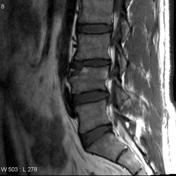
View Limbus Vertebra Mri Findings Pictures. All patients shared loss of disk height, altered disk hydration and variable herniation of nuclear material. Posterior limbus vertebra have been reported to cause nerve compression.
Limbus Vertebra Radiology Reference Article Radiopaedia Org from prod-images-static.radiopaedia.org
Bone spect images revealed significantly increased tracer activity in the region of the anterior l4 vertebral body, which was shown on mri to. Limbus vertebra is defined as the presence of an ossicle or an adjacent bone affecting the margin angle of the vertebral bodies. Shades of gray matter the routine mri is presented as black and whit…
Limbus vertebra develops in the immature skeleton due to focal herniation of the disc through the vertebral endplate, isolating fragment of the peripheral secondary ossification center (ring apophysis).
All patients shared loss of disk height, altered disk hydration and variable herniation of nuclear material. It is not associated with acute trauma. The image on the left shows a t2 mri of a normal brain. Limbus vertebra refers to the herniation of the disc under the ring apophysis before it fuses with the vertebral body.